Anticipations and Significance in Medical Diagnosis for Vascular Ultrasound Diagnostics
Vascular Ultrasound Diagnostics Benefits
Has your doctor prescribed a vascular ultrasound? This may sound intimidating, but it is actually painless and easy to prepare for, and requires no downtime. It is also a very effective diagnostic tool, helping doctors to evaluate blood flow, identify issues, and recognize vascular complications. Vascular ultrasound diagnostics can also help track a patient’s progress after treatment or a procedure. Some of the reasons doctors recommend vascular ultrasounds are to identify the source of leg swelling, leg pain, or signs of venous insufficiency. They can also use vascular ultrasound to detect plaque buildup.
What is a Vascular Ultrasound?
This non-invasive, safe procedure uses sound waves to create images of blood vessels. By reflecting off of blood cells moving within the blood vessels in the body, sound waves can allow a physician to precisely assess the movement and speed of the cells, along with the condition of the blood vessels. Using a vascular ultrasound physicians can determine blood flow to specific organs and tissues, and can detect blockages and abnormalities within the blood vessels. They can check to see if a blood vessel is enlarged, examine varicose veins, and much more. A vascular ultrasound can help a doctor determine whether angioplasty is necessary, and can assess the success of a surgical procedure.
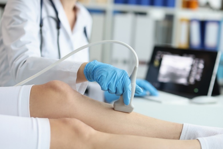
What Can a Patient Expect During an Ultrasound?
The ultrasound process is non-invasive and requires no preparation. The patient’s only responsibility is to lie on an exam table and cooperate with instructions. Depending on which area is to be assessed, the patient may be asked to lie on his or her side or back. The sonographer will apply thick gel to the patient’s skin and use a transducer on the area to be examined. A transducer is a handpiece that glides across the skin and creates sound waves. These waves echo off of the body tissue, and these waves are turned into images by a computer. Vascular ultrasounds do not hurt, and the sonographer will glide the handpiece at various angles, to thoroughly capture the desired images. Sometimes, ultrasounds are used to aid a physician with surgery or another procedure. When a vascular ultrasound is performed at that time, a local anesthetic will often be provided because of the pain caused by the procedure. Once the ultrasound is complete, the sonographer will wipe away the gel, and the patient will be free to go.
Why a Vascular Ultrasound?
Ultrasounds are valuable tools for vascular specialists. They provide information that guides the doctor in creating an effective treatment plan. Ultrasound images help them evaluate varicose veins and use the right treatment strategy, Identify blood clots or heavy plaque, and evaluate the blood supply. Vascular ultrasounds can also be used during treatment, to help guide a doctor as a needle is placed into a blood vessel, for example. They are extremely effective when used for diagnostic purposes, because they allow for the diagnosis of several different conditions, including:
- Chronic Venous Insufficiency: This condition happens when the veins have difficulty getting blood from the limbs back to the rest of the body. This can result in blood pooling in the legs or flow backwards, causing varicose veins.
- Peripheral Artery Disease (PAD): This common circulatory problem narrows arteries, preventing oxygen-rich blood from reaching the body’s limbs. This is often the result of atherosclerosis and can cause painful muscle cramps.
- Carotid Artery Disease: Similar to peripheral artery disease, CAD affects the carotid arteries on both sides of the neck. When the blood supply in these arteries is limited, it reduces the amount of blood sent to the brain, and can result in a stroke, in severe cases.
- Deep Vein Thrombosis: This is a very serious condition in which a blood clot forms in a large, deep vein, typically in the leg. If that clot breaks loose, it can lodge in the lungs, causing a pulmonary embolism, which can potentially be fatal.
For Vascular Ultrasounds and High Quality Care, Contact Arizona Vein & Laser Institute
If you’re seeking diagnosis or treatment for any vascular or cardiovascular issue, trust the board-certified physicians at the Arizona Vein & Laser Institute. Using the most advanced technology, the vascular and cardiovascular surgeons at the Arizona Vein & Laser Institute provide care for all types of venous diseases. With over 40 years of experience, our team of experienced physicians can devise the right treatment plan to address your venous disease problems. For more information contact us through our website.